 english

Ostatnia modyfikacja podstrony: 14.04.2023 12:42
CAVRI Datasets for OCT Retina Image Analysis
We have made available the world's first annotated SD-OCT dataset from subjects with vitreomacular traction (VMT) and vitreomacular adhesion (VMA). In order to receive free-of-charge access to the dataset, please fill out the appropriate form (to be downloaded below), and send it via e-mail with the subject "DATASET ACCESS REQUEST" to: agnieszka.stankiewicz@put.poznan.pl and tomasz.marciniak@put.poznan.pl.
Datasets:
1. CAVRI-A dataset (Preretinal space segmentation)
This database contains 50 (25 subjects with VMA and 25 subjects with VMT) 3D scans with PCV, ILM and RPE annotations. The images were gathered with the use of Avanti RTvue device with 3D Retina scanning protocol (volumetric scan with 141 cross-sections - 7050 B-scans in total).
| Data | Description | Download |
|---|---|---|
| OCT data (.tiff) |
anonimized 50 folders, each with 141 OCT B-scan cross-section (3D scan, 7x7 mm, 640x385 px) |
[download] |
|
segmentation data (.json) |
.json file with struct containg vectors for 10 borders: PCV, ILM, RNFL/GCL, IPL/INL, INL/OPL, OPL/ONL, BMEIS, IS/OS, IB_RPE, OB_RPE. Each vector stores data for 141 B-scans, 385 pixel positions. | [download] |
Data request form: [download_A_form]
Code for preretinal space segmentation developed for the paper "Segmentation of Preretinal Space in Optical Coherence Tomography Images Using Deep Neural Networks" (listed below) can be downloaded from the github repository: https://github.com/krzyk87/pcv_segmentation.
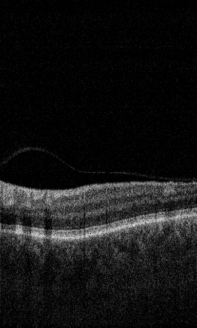
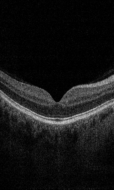
Example images:
| OCT B-scan |
Illustration of segmented regions |
|---|---|
|
|
shades from the top: |

Papers:
Stankiewicz A., Marciniak T., Dąbrowski A., Stopa M., Marciniak E., Obara B., Segmentation of Preretinal Space in Optical Coherence Tomography Images Using Deep Neural Networks, Sensors (Special Issue: Biometric Technologies Based on Optical Coherence Tomography (OCT)), vol. 21, no. 22, 7521, DOI: 10.3390/s21227521, 2021, https://www.mdpi.com/1424-8220/21/22/7521
Stankiewicz A., Marciniak T., Dąbrowski A., Stopa M., Rakowicz P., Marciniak E., Denoising methods for improving automatic segmentation in OCT images of human eye, Bulletin of the Polish Academy of Sciences, Technical Sciences, vol. 65, no. 1, pp. 71-78, DOI: 10.1515/bpasts-2017-0009, 2017, https://journals.pan.pl/dlibra/publication/121296/edition/105686/content
Stankiewicz A., Marciniak T., Dąbrowski A., Stopa M., Rakowicz P., Marciniak E., Improving Segmentation of 3D Retina Layers Based on Graph Theory Approach for Low Quality OCT Images, Metrology and Measurement Systems, vol. 23, no. 2, pp. 269-280, DOI: https://doi.org/10.1515/mms-2016-0016, 2016, https://journals.pan.pl/dlibra/publication/104499/edition/90412/content
2. CAVRI-B dataset (Preretinal space parameterization)
This database contains 104 (12 subjects with VMA and 14 subjects with VMT) 3D scans with PCV, ILM and RPE annotations. Each subject was examined 4 times in the course of 3 to 4 years of observation. The images were gathered with the use of Avanti RTvue device with 3D Retina scanning protocol (volumetric scan with 141 cross-sections).
- OCT data (.tiff)
- segmentation data (.xml / .json / .mat)
- database statistics (.xlsx),
Papers:
Stopa M., Marciniak E., Rakowicz P., Stankiewicz A., Marciniak T., Dąbrowski A., Imaging and Measurement of the Preretinal Space in Vitreomacular Adhesion and Vitreomacular Traction by a New Spectral Domain Optical Coherence Tomography Analysis, Retina, 37(10), pp. 1839-1846, doi: 10.1097/IAE.0000000000001439, PMID: 28045789, 2017, https://journals.lww.com/retinajournal/.
Stankiewicz A., Marciniak T., Dąbrowski A., Stopa M., Marciniak E., "A New OCT-based Method to Generate Virtual Maps of Vitreomacular Interface Pathologies", 18th International IEEE Conference on Signal Processing Algorithms, Architectures, Arrangements and Applications (SPA 2014), Poznań, Poland, pp. 83-88, 2014, https://ieeexplore.ieee.org/document/7067275
3. CAVRI-C dataset (Vessels segmentation)
This database contains 24 3D scans with ILM, NFL/GCL, GCL/IPL, IPL/INL, INL/OPL, IS/OS and RPE layers annotations for 24 healthy subjects in the average age of 27. The images were gathered with the use of Avanti RTvue device with 3D Retina scanning protocol (volumetric scan with 141 cross-sections). This dataset contains also manual retina vessels segmentations for the fundus reconstructed image.
| Data | Description | Download |
|---|---|---|
| OCT data (.tiff) |
anonimized 24 folders, each with 141 OCT B-scan cross-section (3D scan, 7x7 mm, 141 images - each 640x385 px) |
[download] |
|
segmentation data (.xml / .json / .mat) |
||
| fundus reconstructions (.bmp) |
3 variants of reconstruction method (see papers listed below)
- P1 reconstruction (141x385 and 385x385) |
[download] |
Data request form: [download_C_form]
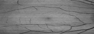
Example images:
 |
P1 reconstruction:
P3 reconstruciton:
Reference: |
| OCT B-scan |
Reconstructed fundus |


Papers:
Marciniak T., Stankiewicz A., Dąbrowski A., Stopa M., Rakowicz P., Marciniak E. Measurement of retina vessels by segmentation of images reconstructed from optical coherence tomography data. Metrology and Measurement Systems, 26(3), 449-461, 2019, https://journals.pan.pl/Content/113100/PDF/01_MMS_3_INTERNET.pdf
Marciniak T, Stankiewicz A, Zaradzki P. Neural Networks Application for Accurate Retina Vessel Segmentation from OCT Fundus Reconstruction. Sensors, 23(4):1870, 2023, https://doi.org/10.3390/s23041870 https://www.mdpi.com/1424-8220/23/4/1870
DISCLAIMER
All images included in this website have been fully de-identified. Any dates associated with the imaging files do not relate to the subject or to date of image acquisition. Images are intended for use in research and educational settings.
Commercialization/redistribution of the images is prohibited.
In the unlikely event that you identify any remaining identifiers in the images, you are prohibited from further disclosure and should destroy all copies of the image and immediately notify the owner of this webpage at: tomasz.marciniak@put.poznan.pl or agnieszka.stankiewicz@put.poznan.pl.
All use of the images should include citation and credit to their corresponding paper.